Where Microscopy Meets Discovery
Trusted by more than 50 world-leading research labs.


SPATIAL TM
Multiplex image analysis right here in your web browser.
World-class segmentation and registration. Neuroscience toolkit. For multi-channel Immunofluorescence, spatial proteomic, transcriptomics and more.
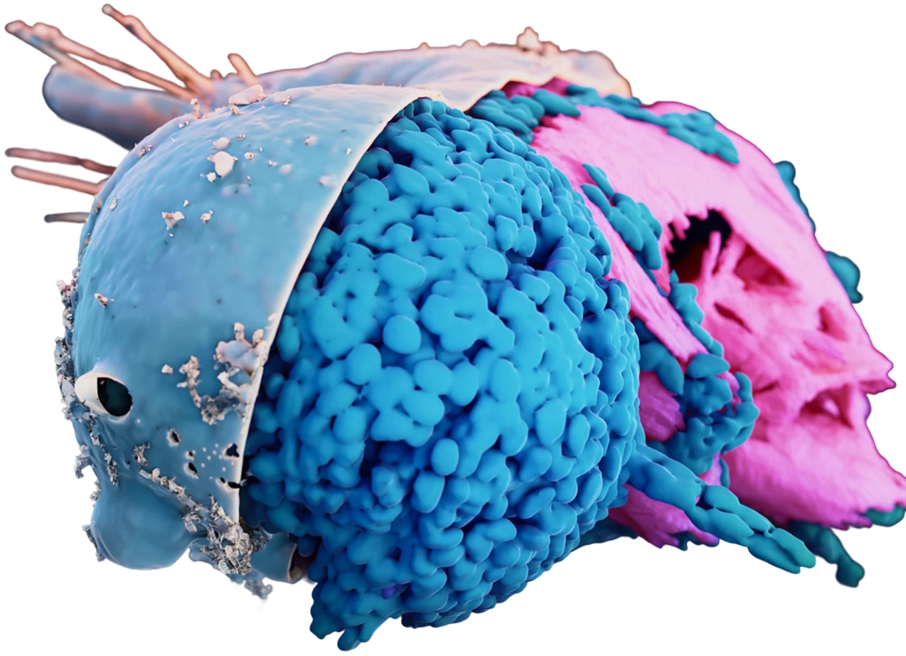
3dEMtrace
Reconstruct serial electron microscopy and tomography volumes.
Reveal cellular structure in unprecedented detail. Map mitochondria, synapses, nuclei, ER and more.
LMtraceTM
Neuron reconstruction made easy
LMtrace is our worry-free pipeline for reconstruction of neurons and glia cells in light microscopy datasets. Just upload your dataset, and we take care of the rest!
applications
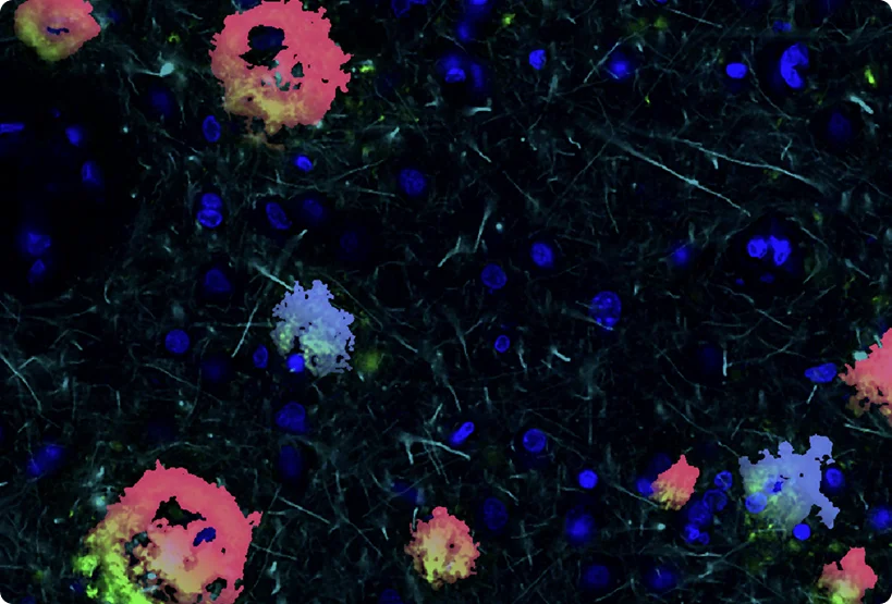
Comprehensive in silico tissue characterization in Alzheimer's and Parkinson's disease by spatial proteomics
Learn how ariadne.ai's cloud-based SPATIALTM platform helps discover spatial signatures that differentiate health from disease in CNS tissue. This application note walks you through neuron and glia segmentation at single-cell resolution from diverse markers (NeuN, IBA1, GFAP) and classification of pathological protein aggregates (including p-Tau, α-Synuclein, TDP-43 and Amyloid β).
Learn more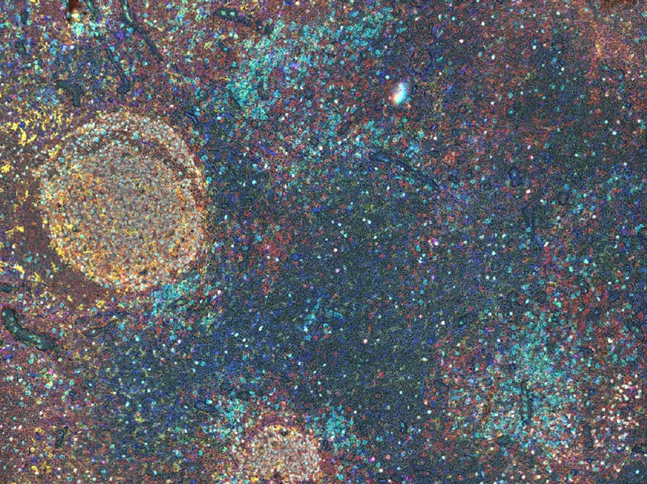
Webinar: High-plex immunohistochemistry - Spatial biology links patient survival with lymph node B cell responses in head & neck cancer
Dr. Ferdinando Pucci discusses his development of a high-plex immunohistochemistry (IHC) protocol and its applications in cancer biology. Learn how SPATIALTM corrects complex tissue deformations with its elastic registration workflow and allows researchers to discover spatial biomarkers subsequent to marker-based phenotyping.
Learn more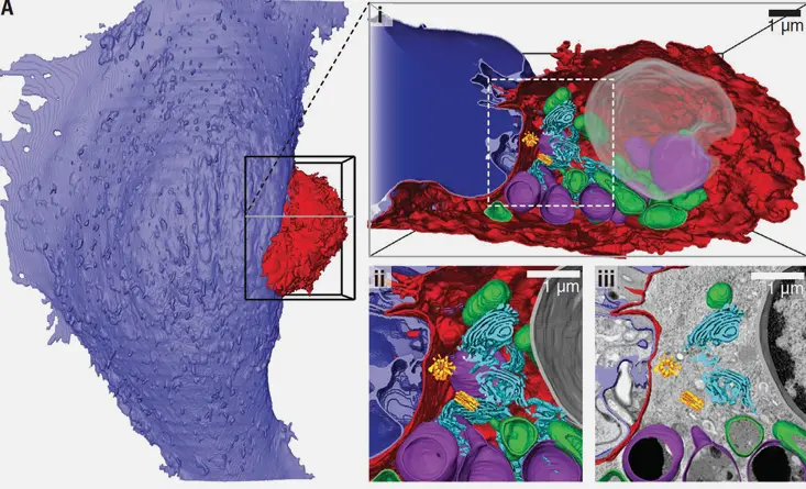
Scalable segmentation in volume electron microscopy (vEM)
Learn how to leverage ariadne.ai's 3dEMtrace(TM) service to scalably and reliably segment diverse structures from vEM, such as cells, synapses, endoplasmic reticulum, mitochondria, Golgi apparatus and much more.
Learn more
Webinar: Identification of microenvironment-dependent myeloid cell heterogeneity in AD with spatial biology
Dr. Bahareh Ajami discusses her research on myeloid cells in Alzheimer's disease using SPATIALTM, highlighting precise cell segmentation, subcellular proteomic analysis, and microenvironment-level insights into neuropathology.
Learn more
Webinar: Decoding Neurodegeneration
Dr. Birgitt Schuele discusses how high-plex spatial proteomics and spatial biology are advancing neuroscience discovery and translating spatial insights into breakthroughs in brain science.
Learn moreMeet Us
Selected Publications
MoreOver the years our services and deep-learning techonology have been used for many high-profile scientific publications. Below you find a collection of image analysis use cases or visit our News site to learn more about recent publications and conference contributions.
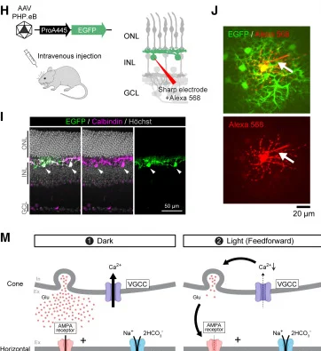
The role of the sodium-bicarbonate cotransporter Slc4a5 in retinal feedback mechanisms
Morikawa et al., Neuron 2024
Learn more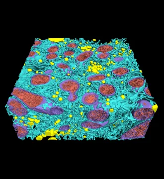
Regulation of liver subcellular architecture controls metabolic homeostasis
Parlakgül et al., Nature 2022
Learn more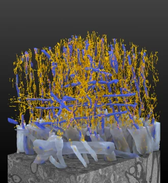
Transverse endoplasmic reticulum expansion in hereditary spastic paraplegia corticospinal axons
Zhu et al., Human Molecular Genetics 2022
Learn more
ESCRT-mediated membrane repair protects tumor-derived cells against T cell attack
Ritter et al., Science 2022
Learn more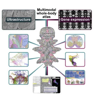
Whole-body integration of gene expression and single-cell morphology
Vergara et al., Cell 2021
Learn more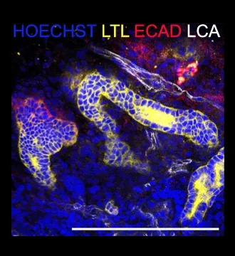
Vasculogenesis in kidney organoids upon transplantation
Koning et al., npj Regenerative Medicine 2022
Learn moreWhat our clients say

Dr. Ferdinando Pucci
Assistant Professor, OHSU

Scientifically excellent
They supported our lab's immuno-oncology work from start to finish. We had some challenging data for them, and they tuned their registration and segmentation to handle it perfectly.

Dr. Oliver Braubach
Director of R&D, Canopy Biosciences
Great segmentation
Their neuron and glia segmentation is the best I've seen yet.
Dr. Bahareh Ajami
Assistant Professor, OHSU
Super responsive
The ariadne.ai team was always there to handle our custom requests quickly and with great scientific rigor. Their neuron and glia segmentation is fantastic.

Dr. C.Shan Xu
Professor of Cellular & Molecular Physiology, Yale School of Medicine
Beautiful segmentation
Ariadne.ai has repeatedly and consistently produced beautiful, top-notch segmentations on the 3D data we generated.

Dr. Anton Arkhipov
Investigator, Allen Institute
Great Experience
Everyone has been very helpful and it's been a great experience working with them. Ariadne has been a great partner in this project.