

Most cell segmentation tools find the nucleus, expand it and stop there.
That is fine for counting cells. It is not fine if you want to understand what those cells are doing.
A paper published today in Nature Neuroscience makes this point precisely. Using spatial proteomics on human Alzheimer’s disease brain tissue, the authors around Bahareh Ajami & Dr. med. Dennis-Dominik Rosmus profiled 704,706 cells across 8 AD patients and 8 healthy controls.
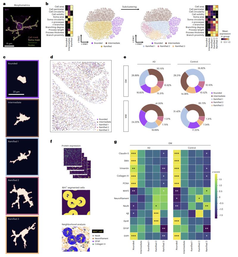
They clustered microglia by protein expression and found subpopulations. Then they added morphology. Then they added spatial neighborhood.
Each layer revealed something the previous one missed.
The key finding, a previously undescribed microglial subpopulation they named human plaque-associated microglia, enriched specifically in AD brains and strongly associated with dense amyloid-beta plaques was invisible to protein expression clustering alone. It only emerged when the spatial context of each cell was taken into account. And that spatial context required knowing where the cell actually was, how large its soma was, how many processes it extended, how it related to surrounding structures.
You cannot get that from a nucleus mask.
Studies like this used to require assembling a pipeline from multiple tools, custom scripts, and the time to learn and maintain all of it. That is time most labs do not have, and expertise most biologists should not need to acquire just to get to the biology.
Here, analysis was done in SPATIAL – one platform, no coding, from raw images to analysis-ready results.
Image depicts figure 4 from the original publication: https://lnkd.in/dwnjhxBW