

SPATIALTM 2
Spatial biology image analysis, from raw slide to result. In your browser.
Alignment, segmentation, phenotyping, QC and spatial cohort analysis in one place, for multiplex immunofluorescence, spatial proteomics and transcriptomics. Nothing to install, no compute to manage, no scripting required.
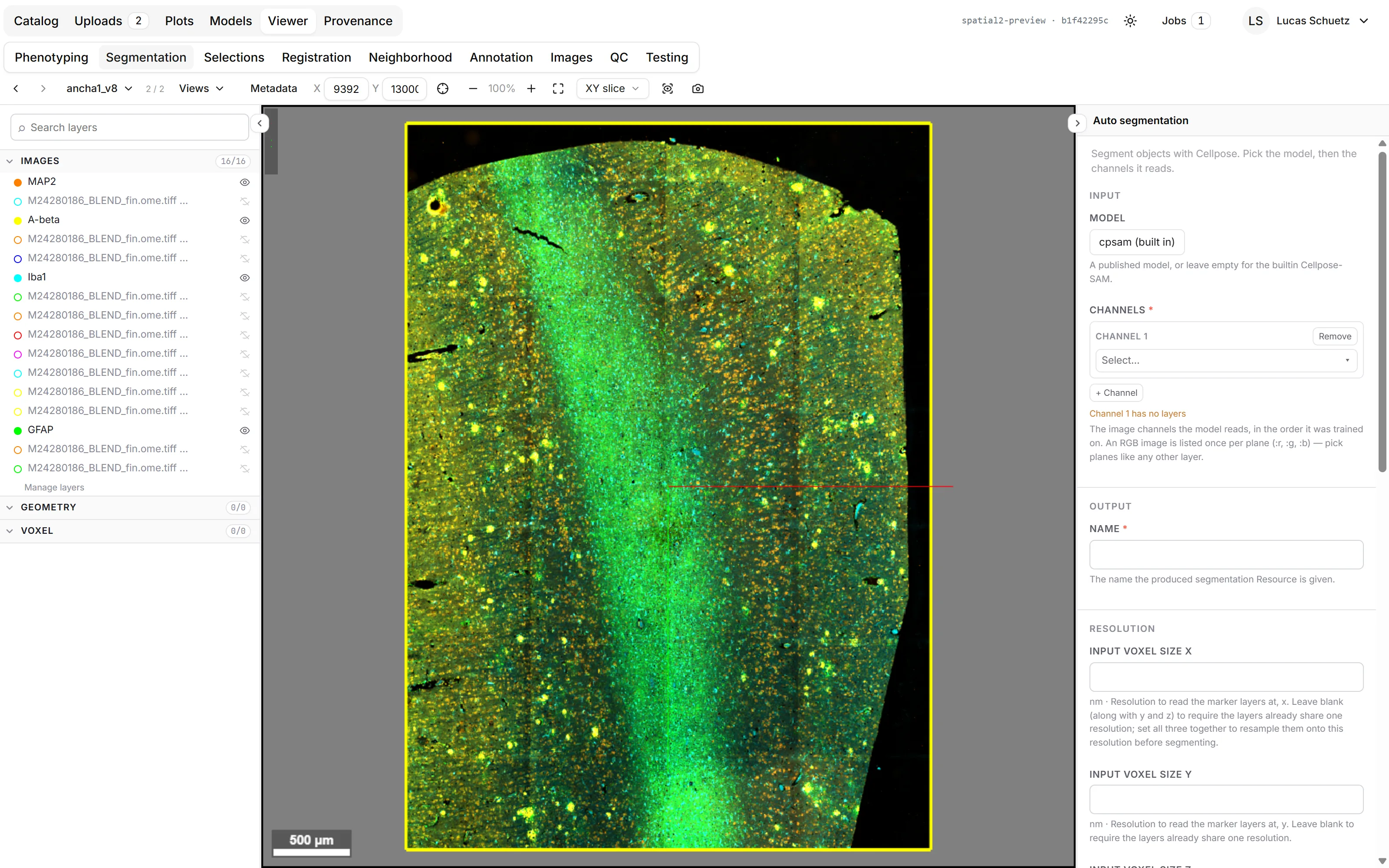
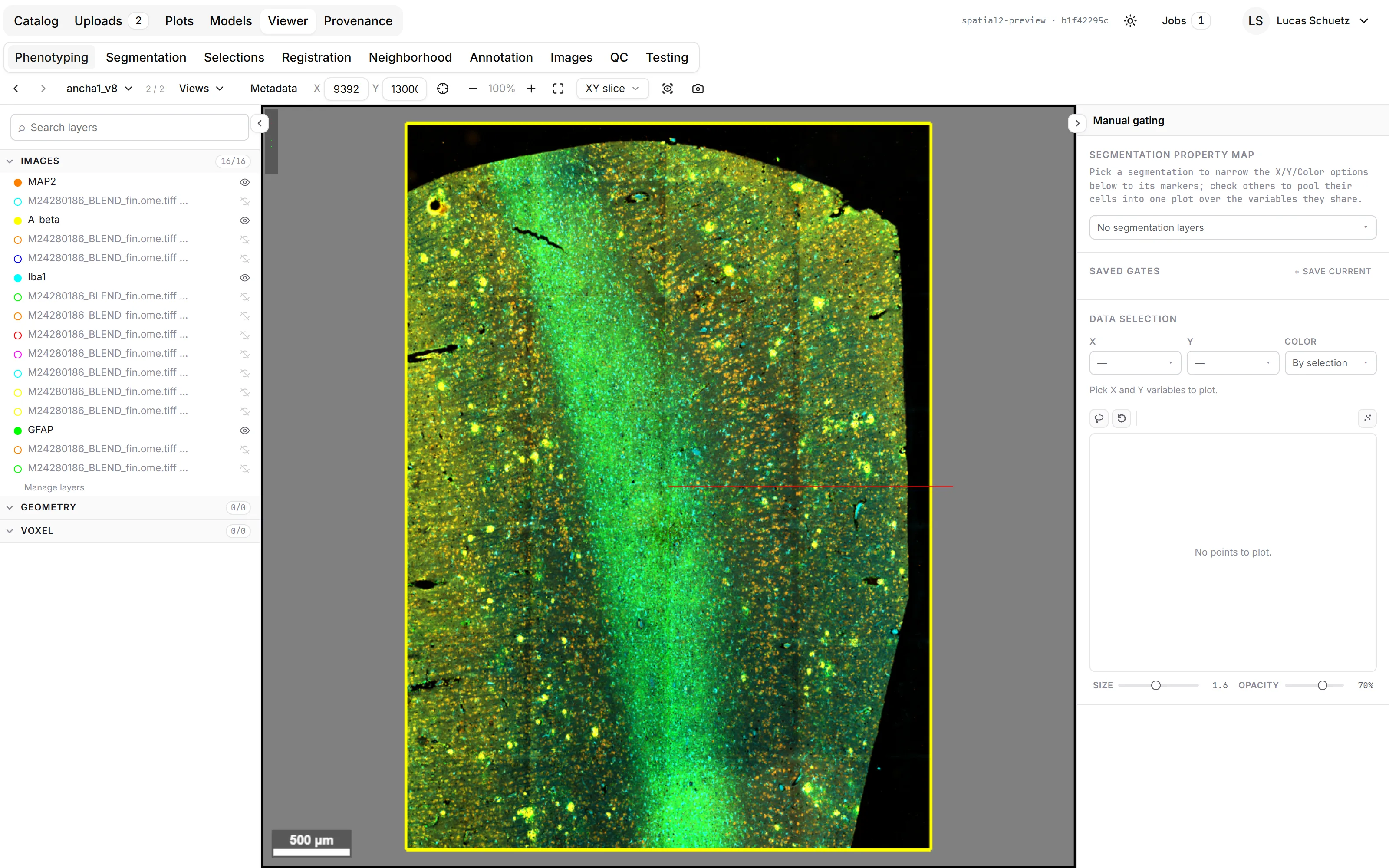
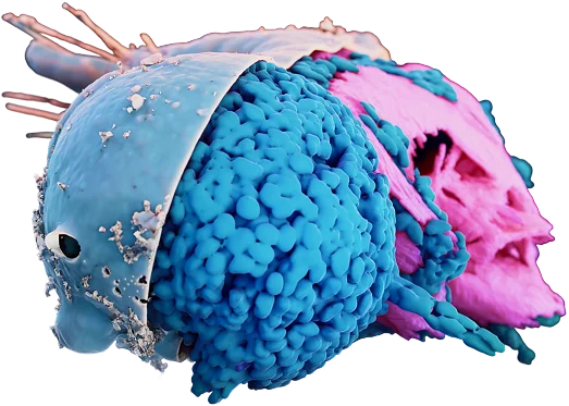
World-class cell segmentation.
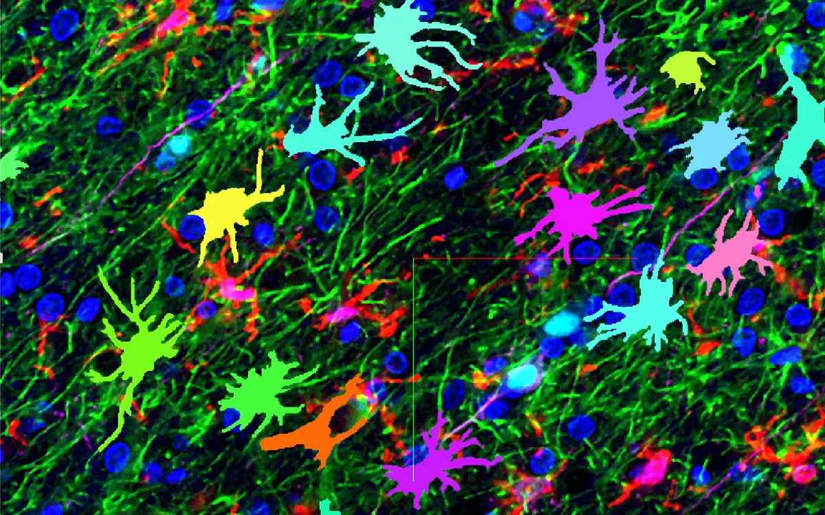
Also for cells with complex morphologies such as neurons and glia. Supports CD45, NeuN, Iba1, GFAP, DAPI and many more.
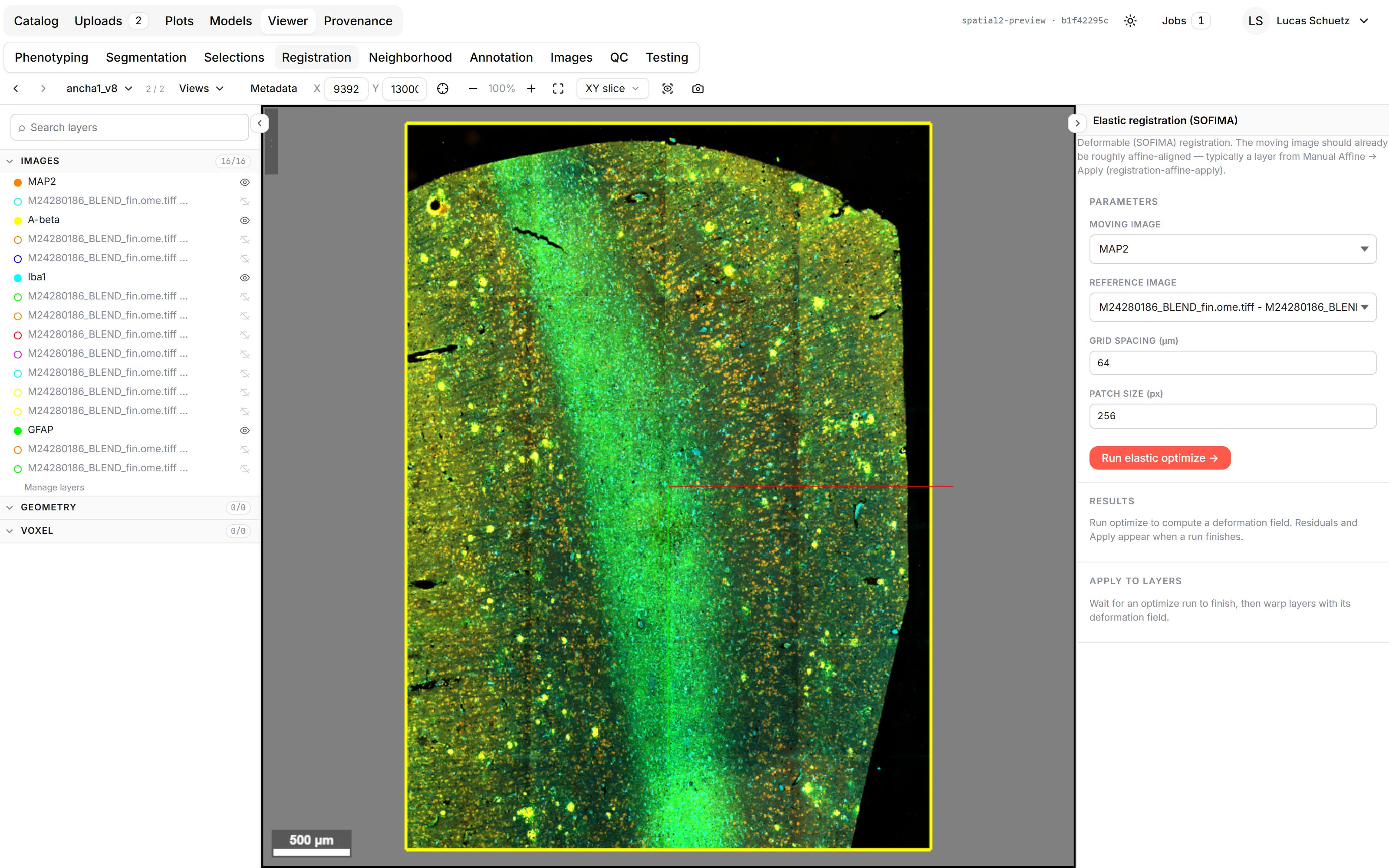
Elastic registration.
Correct complex deformations across consecutive staining cycles, protocols or physical sections.
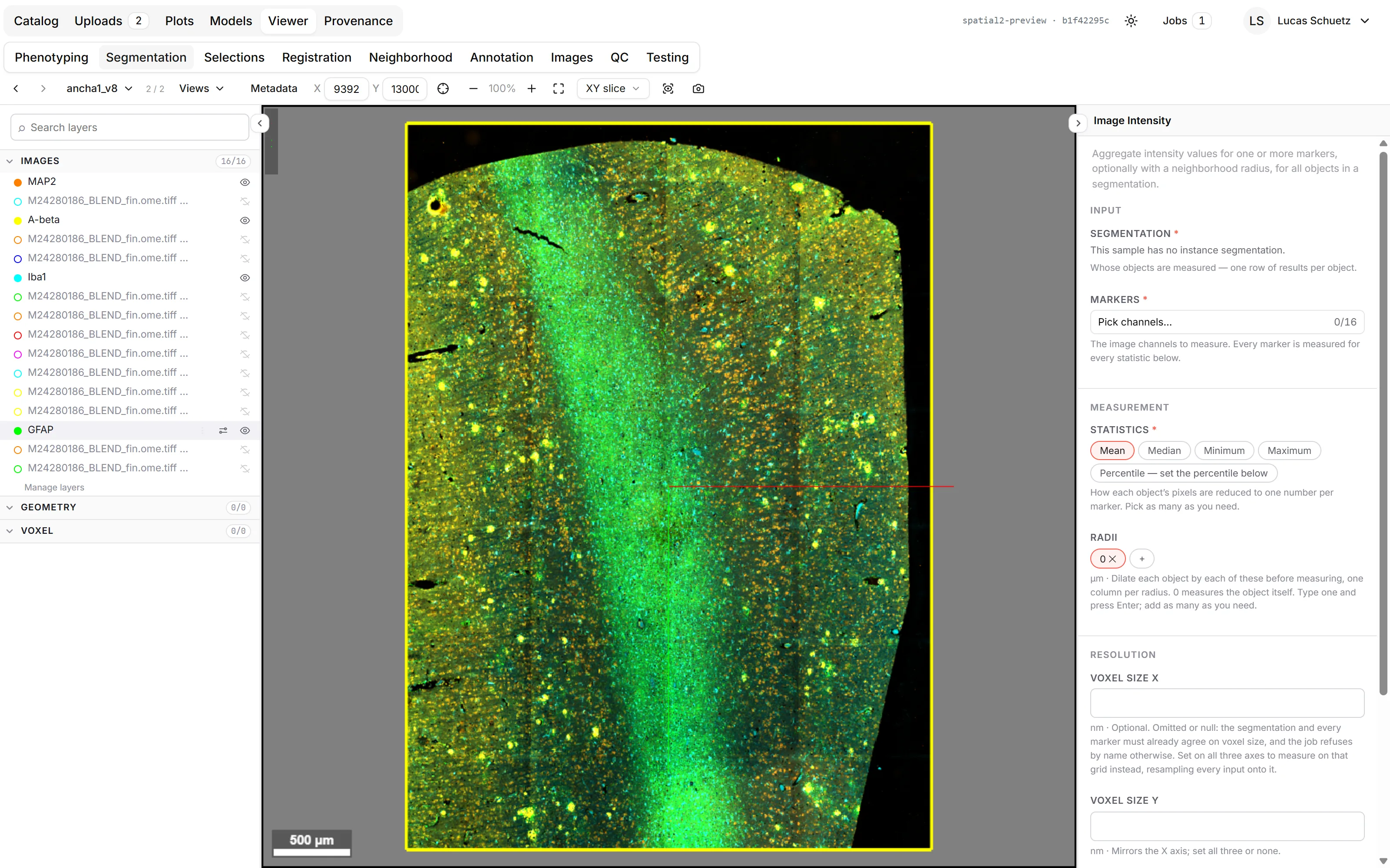
Neuropathology analysis.
Classify pathological protein aggregates of amyloid-β, phospho-tau, α-synuclein, TDP-43 and more.
Segment H&E images.
Leverage well-established IHC protocols, multiplexed.
See it live
Explore SPATIALTM 2 below. A live demo where you can navigate and experiment yourself is coming soon.

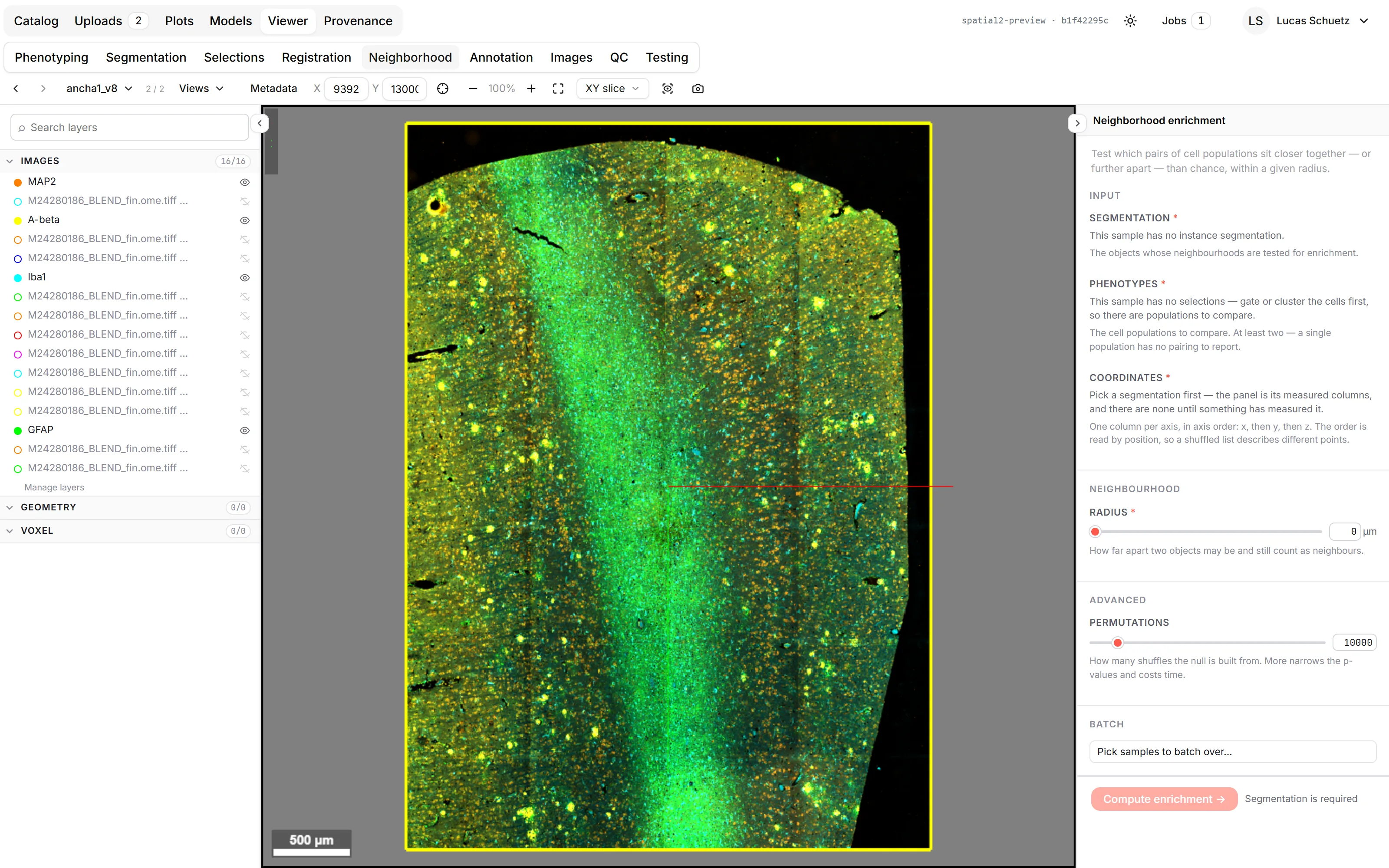
Try SPATIALTM 2 on your own data
These are the real screens of SPATIALTM 2. The clips are recordings of the application, played at one and a half times speed.
Live demo coming soon
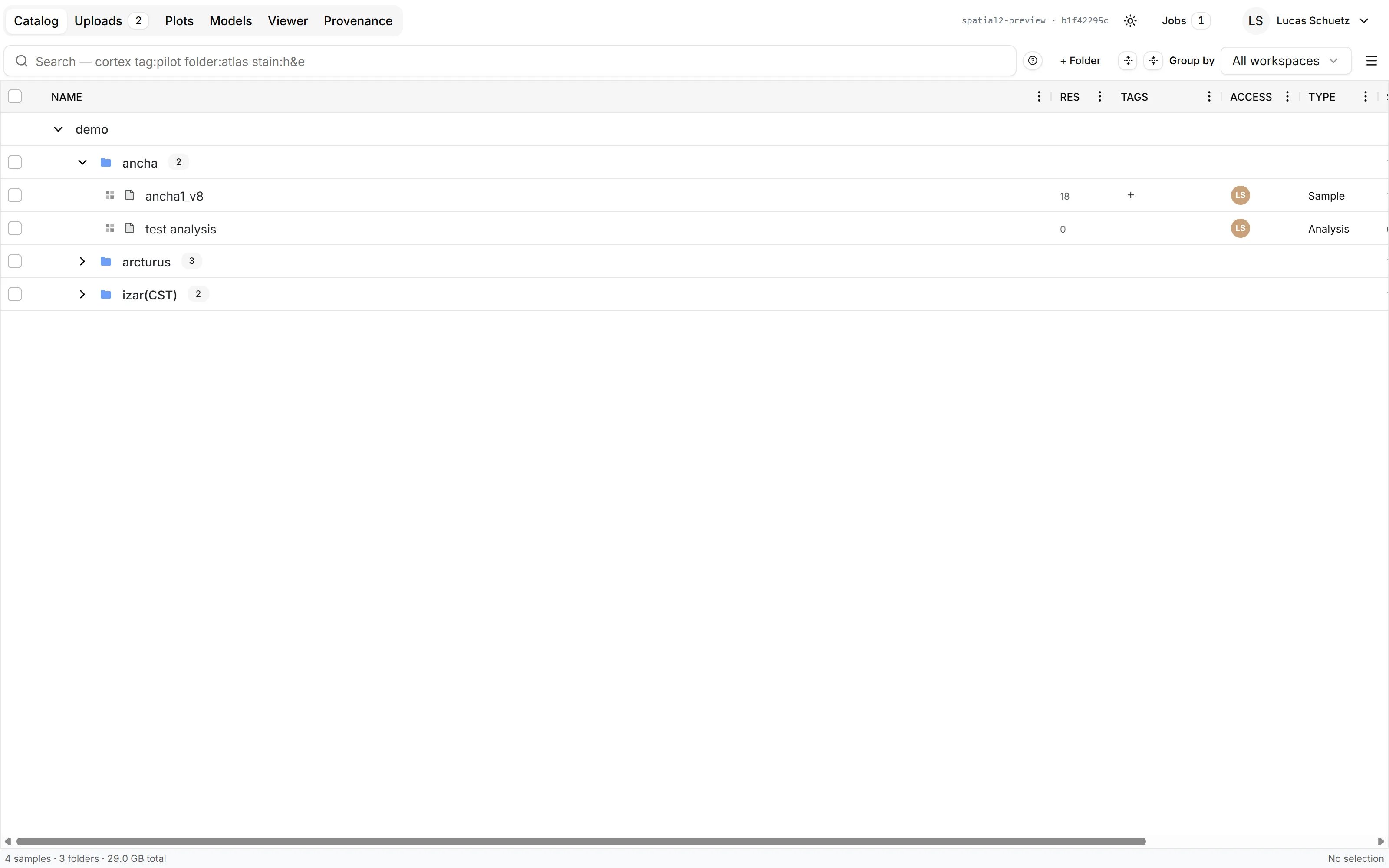
Your whole catalogue in one place. Folders carry their panels and tags with them, so a cohort stays organised as it grows.
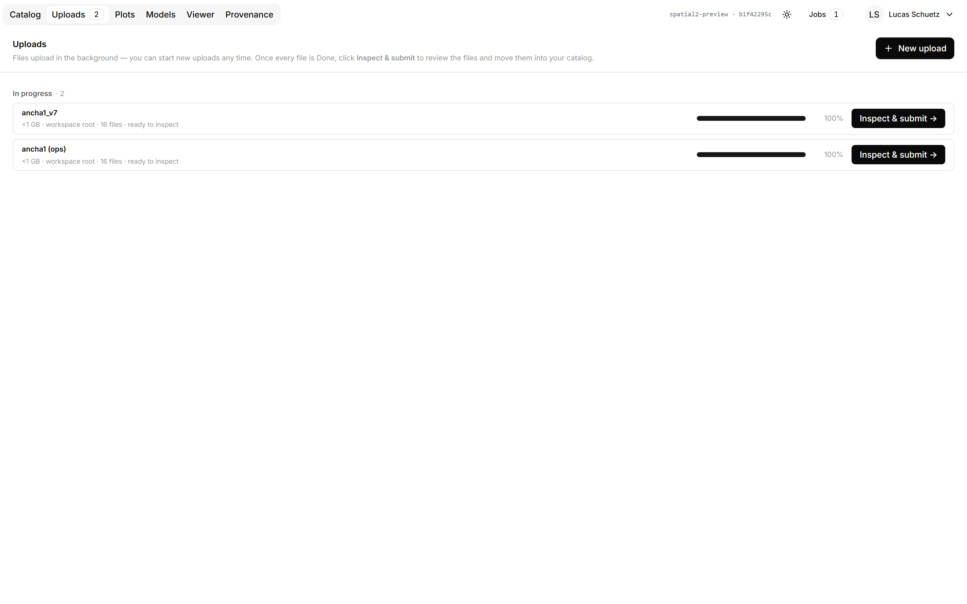
Upload in the background. Drop files in and keep working. Failed uploads can be resumed, and a sample appears in the catalogue once it is complete.
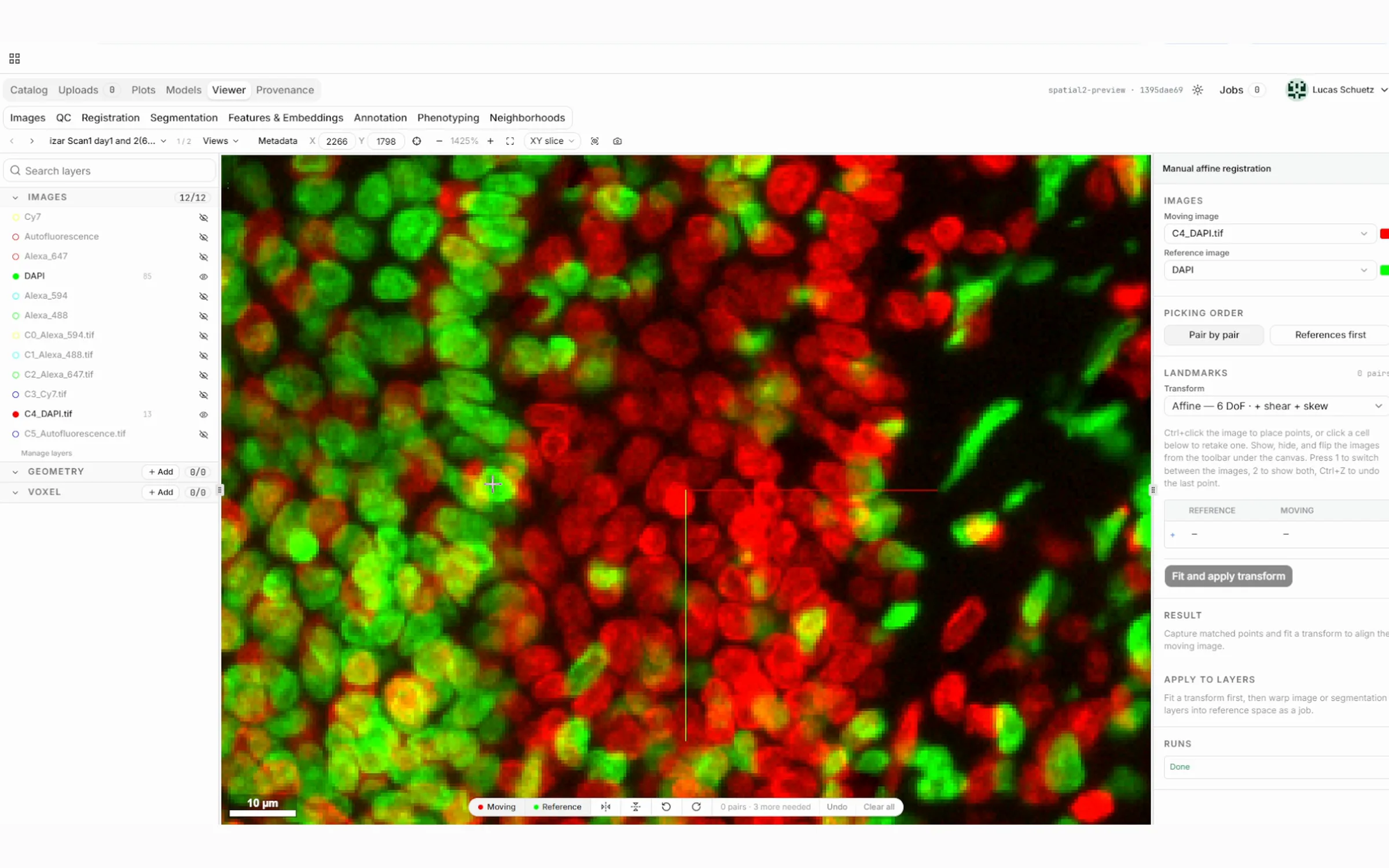
Put the modalities in one frame. Three matched points set the affine transform between any two images, from one section or from several.
Correct the local deformation. Sectioning, hydration and shearing deform tissue locally. The elastic transform follows that, at any image size, and a 3D dataset can be built from consecutive sections.
Segment from the markers that outline the cell. Pre-trained models for neurons, glia and other cell types keep the morphology that matters.
Train directly in SPATIALTM 2. Optimise our models on your own data, or use our training service. Every run keeps its loss curves and its accuracy, so you always know which model produced a result.
Every cell carries its own set of properties. Proteomic, transcriptomic, IMC and morphological properties are measured per cell, so each one carries a unique combination.
Gate, cluster, or both. Automatic cell clustering does the coarse work, then manual gating lets your own experience decide where the populations end.
Find the microenvironments. Recurrent neighbourhoods, neighbourhood enrichment and other jobs find local niches and ligand-receptor interactions.
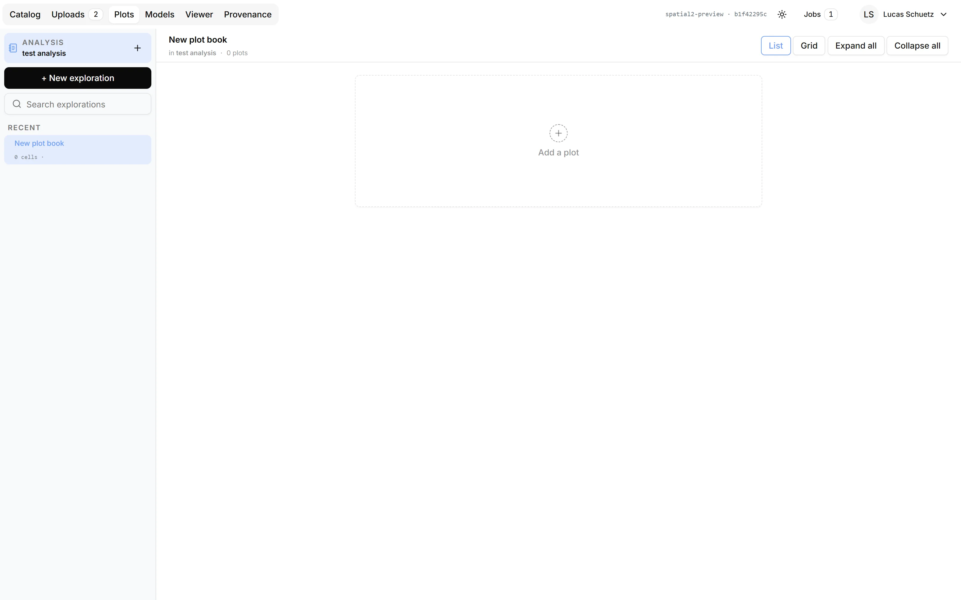
Plot books instead of screenshots. Figures stay attached to the analysis, regenerate when the data changes and always link back to their raw data.
One workflow, from upload to result
Every analysis in SPATIALTM 2 follows the same path, with optional branches for training your own models, multi-omics registration and batch processing.
Explore the features
Upload
Vendor output or open formats, straight from the instrument.
Register
Elastic alignment across cycles, protocols and sections.
Segment
Cells with real morphology: neurons, glia, tumour, immune cells.
Phenotype
Gate, cluster and compare across conditions and regions.
Quantify
Spatial statistics and morphometric biomarkers, exported or shared.
Batch
Any step over a whole cohort, with the same settings on every sample.
Any disease area. First in neuropathology.
SPATIALTM 2 works on any tissue and any panel. It was built with neuropathologists on human brain tissue, and that is where it goes deepest. Hover an image to see the segmentation.
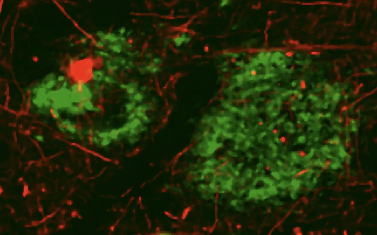
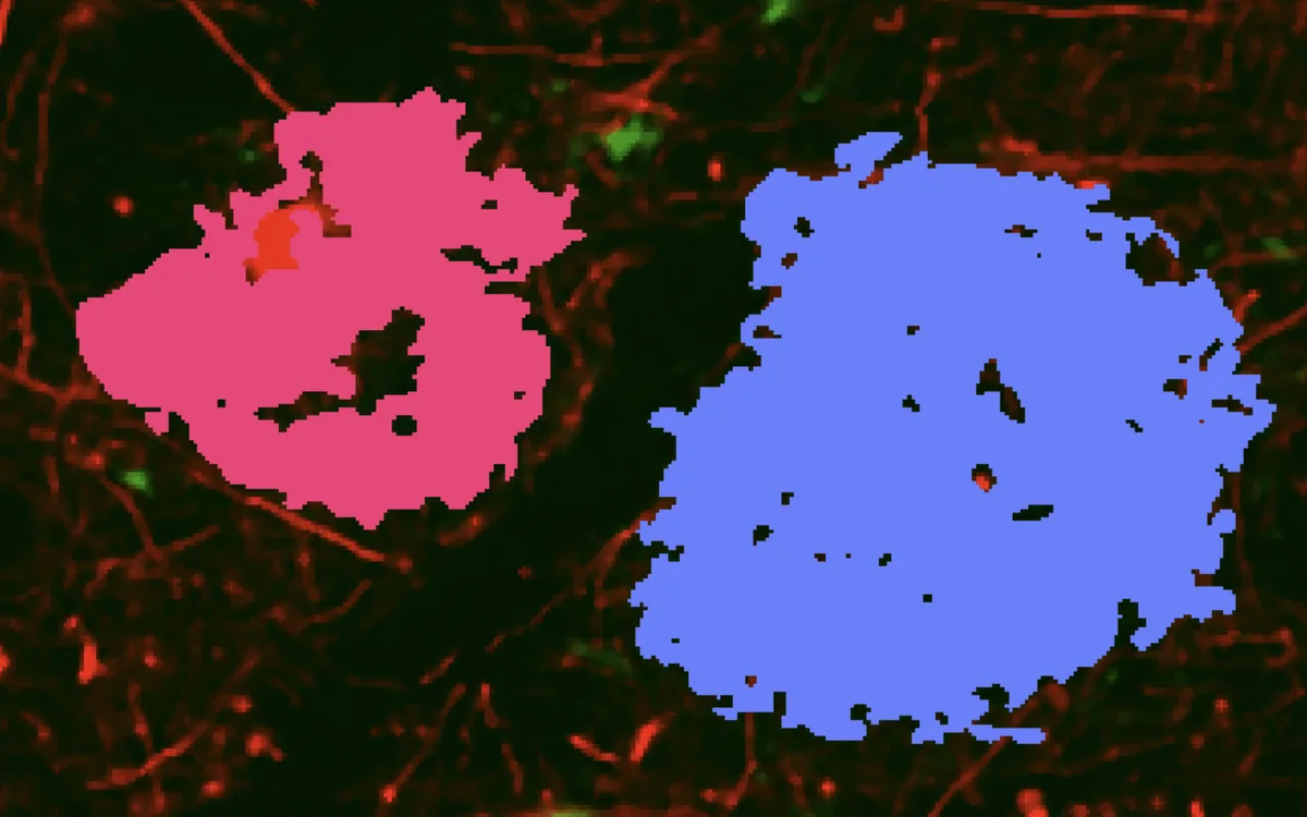
Alzheimer’s disease
Plaques, tangles and the cells around them
SPATIALTM 2 comes with advanced classification algorithms for:
- amyloid-β plaques, dense-core or diffuse
- Phospho-tau, tangles or neuropil threads
- Microglia
- Astrocytes
- Neurons

Parkinson’s disease
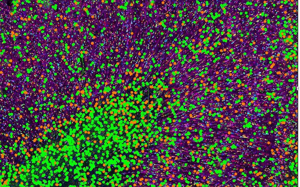
α-synuclein pathology in context
Lewy body pathology is stage dependent. In a TH stain, SPATIALTM 2 segments and classifies:
- α-synuclein aggregates as objects of their own
- Lewy bodies, halo and core
- Dopaminergic neurons
- Microglia and astrocytes around the pathology
 Work in progress
ALS
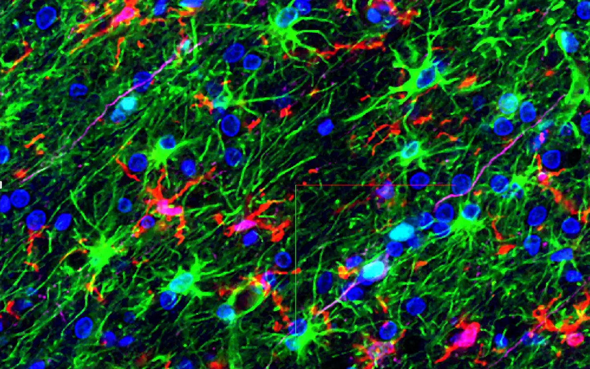
TDP-43 aggregates as objects of their own
In motor cortex and spinal cord, SPATIALTM 2 segments and classifies:
- TDP-43 inclusions, skein-like, round or dystrophic neurites
- Neurons that have lost TDP-43 from the nucleus
- Motor neurons, down to Betz cells
- Microglia and astrocytes
- Myelin, and where it thins along the tract
Glioblastoma
The invasive edge, cell by cell
Across the tumour and its margin, SPATIALTM 2 segments and classifies:
- Tumour cells and the fraction that is proliferating
- Microglia and infiltrating macrophages
- T cells and other immune subsets
- Vessels and the perivascular niche
- Pseudopalisading necrosis at the invasive front
New in SPATIALTM 2
Works with your instrument
Most common file types are supported, and we keep adding more.
Akoya
Phenocycler
Bruker Spatial Biology
CosMx, CellScape
Lunaphore
COMET
10x Genomics
Visium, Visium HD, Xenium
Vizgen
MERSCOPE
Zeiss and Leica
CZI, LIF
Open formats
OME-TIFF, TIFF series
Something else?
Tell us what you run. The list keeps growing.
What researchers say
“They supported our lab’s immuno-oncology work from start to finish. We had some challenging data for them, and they tuned their registration and segmentation to handle it perfectly.”

“Their neuron and glia segmentation is the best I’ve seen yet.”

“The team was always there to handle our custom requests quickly and with great scientific rigor. Their neuron and glia segmentation is fantastic.”